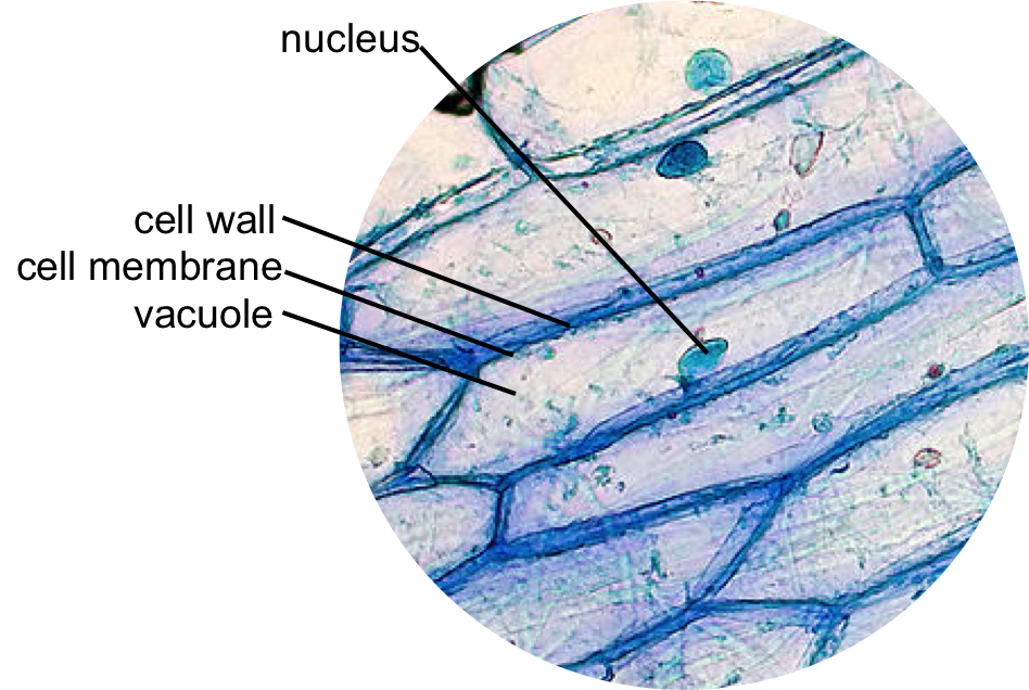
Onion Cell Under Microscope
X100 when printed at. Line up one of the divisions on the eyepiece graticule with a fixed point on.

Epidermal Onion Cells Under A Microscope Plant Cells Appear Polygonal From The Cell Diagram Plant Cell Diagram Plant Cell
4 Place a drop of iodine stain on your onion tissue.

. Onion cells exhibit a brick-like shape under the microscope. It has a cell wall cell membrane cytoplasm nucleus. STEP 1 - Carefully cut an onion in half or.
Onion Cells Under the Microscope Introduction The bulb of an onion is formed from modified leaves. An onion is a multicellular plant. This lab is perfect for an introductory biology course at the high school or college level.
The onion cell is a plant cell that can be obtained by peeling off an onion. An onion a slide and cover slip a cotton bud some food colouring a plate to put the cotton bud on and of course a microscope. While photosynthesis takes place in the leaves of an onion containing.
Add a drop of purple stain specific for animals and cover with a cover. Place a stage micrometer on the stage of the microscope. 3 Place the single layer of onion on a glass slide.
Download all free or royalty-free photos and images. Its intended to be used as a way for students to explore prokaryotic and eukaryotic cell types. Garden onion Bulb Onion Common Onion Allium cepa cell tissue of a garden onion with dyed chromosomes light.
Thus onion being a plant. Up to 10 cash back Light micrograph LM of a transverse section of onion Allium cepa root tip to show cells undergoing mitosis nuclear division. Up to 10 cash back Onion cells under the microscope.
2 put your slide on. Gently roll and rub the toothpick onto the top of a glass slide in an area that will be visible through the microscope. Station remove the single layer of epidermal cells from inner side of the scale leaf.
STEP 2 Place the layer of onion epidermis carefully on the glass slide and cover with a cover slip. The presence of a rigid cell wall and a large vacuole is a characteristic feature of a plant cell. Peel a thin layer of onion the epidermis off the cut onion.
Your Onion Cells Under Microscope stock images are ready. Preparation of onion cell slide and viewing under a light microscope to view cheek cells gently scrape the inside lining of your cheek with a toothpick. Use them in commercial designs under lifetime perpetual worldwide.
Using a microscope to measure cell size.

Onion Epidermis With Large Cells Under Light Microscope Clear Epidermal Cells O Ad Microscope L Microscopic Cells Things Under A Microscope Dna Project

The Famous Onion Skin Cells X 100 Dyed With Iodine

Why Is Iodine Stain Used On Onion Cells Iodine Cell Stain

Epidermal Onion Cells Under A Microscope Plant Cells Appear Polygonal From The Cell Diagram Plant Cell Diagram Plant Cell

Onion Cells Under A Microscope Requirements Preparation Observation Plant And Animal Cells Animal Cell Plant Cell

Onion Cells Google Images Ilustracoes Felipao
0 Response to "Onion Cell Under Microscope"
Post a Comment